The Art of Laboratory Science
Medical laboratory scientist honored at international art competition.
Picture a hospital or clinical laboratory. What do you see? Perhaps lab coats and gloves, test tubes and beakers, petri dishes and instruments. A clean, white, sterile world of medical equipment occupied by busy, dedicated professionals. But if you look closer you will find a world filled with vibrant colors and artistic ambitions.
.JPG)
Laboratory science and art go hand-in-hand according to Meredith Herman, MSU Biomedical Laboratory Diagnostics class of 2017. Herman is currently a post-sophomore pathology fellow at the University of Toledo Medical Center and medical student in MSU’s College of Osteopathic Medicine.
“Medical laboratory science is such a visually oriented specialty in medicine. When you're studying pathology, looking at slides on the microscope, you have to look closely at the details of the tissue. Inspecting features like how cells are spaced in the tumor architecture. Are they forming papillary clusters? Glands? Solid nests? You rely on your pattern recognition to sort the possibilities to get down to what's going on in a patient. So, it's art and medicine, together.”
One of Herman’s recent paintings, “Life Magnified,” which was inspired by her experiences in BLD and the pathology fellowship, received a 'Highly Commended' by the Royal College of Pathologists during its annual international art competition, the Art of Pathology Competition. This year's theme was titled: ‘Pathology: at the heart of healthcare.' Additionally, this painting was featured in the International Pathology Day 2020 program.
It’s the role of the lab to discern what pathology has occurred in a patient and Herman's painting reflects all the different aspects of a hospital laboratory which work together to reach a diagnosis.
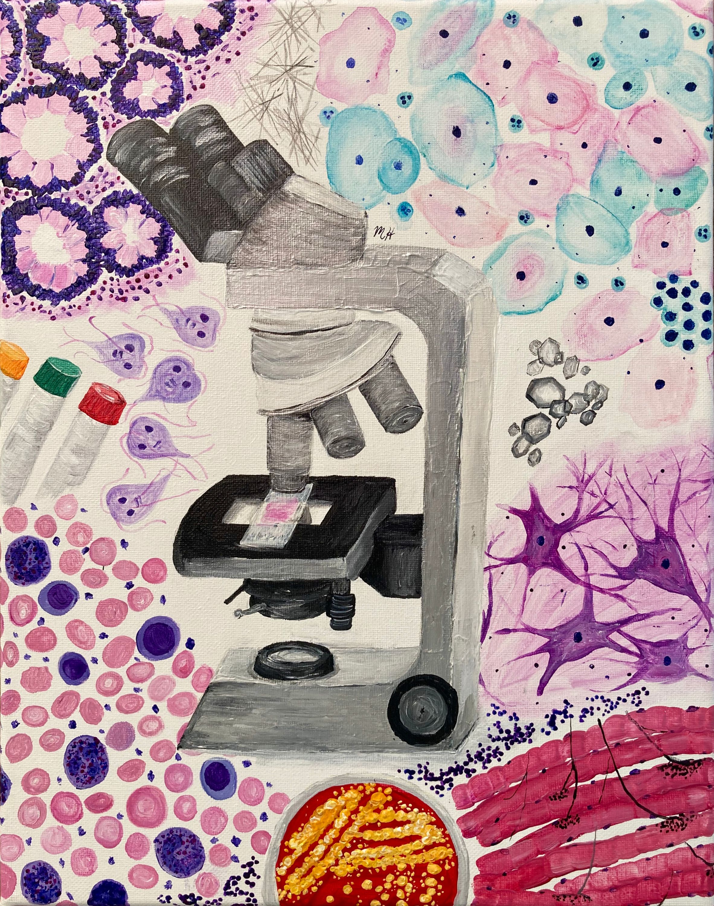
"Laboratory disciplines really are at the center of healthcare," said Herman while explaining her artwork. "In the center of the painting, there's a microscope with a slide on the stage containing a piece of tissue. The images around the microscope contain elements that represent various aspects of the hospital laboratory. At the top left, you see histology of colon. To the right are urine crystals. The top right corner shows a Pap smear from cytology. Beneath that, there's neural tissue. The bottom right corner is skeletal muscle tissue at the neuromuscular junction. The bottom center is a bacterial culture plate with Staph aureus. And the bottom left corner is hematology, where we're looking at blood cells. Above that are test tubes and next to them, the parasite, Giardia."
The result truly illustrates the colorful, captivating cellular world of laboratory medicine.
Herman is not alone in seeing the beauty of the laboratory. Nicole Vezina, BLD ‘21, echoes the connection of art and lab science.
“Laboratorians need to know color," said Vezina. "With spectrophotometry in particular, color changes in an assay indicate whether it's positive or not, which is critical for a patient's life.”

Vezina channeled her creativity through drawing during the stay home, stay safe restrictions. She started a REDBUBBLE shop where she promotes laboratory professionals by selling her artwork as stickers.
“Laboratory sciences are just coming to the surface," Vezina noted. "More people are more aware of how many professions exist behind just nurses and doctors. I wanted to depict all the different kinds of women and all the different kinds of people who are working behind the scenes. I made the laboratory stickers with a bunch of different cultures and different types of women so that they could see themselves in their profession and be able to express themselves.”
Overlaps of art and lab sciences, for both Herman and Vezina, extend down to the foundation of their studies. Both, equipped with notebooks filled with illustrations, learn through drawing.
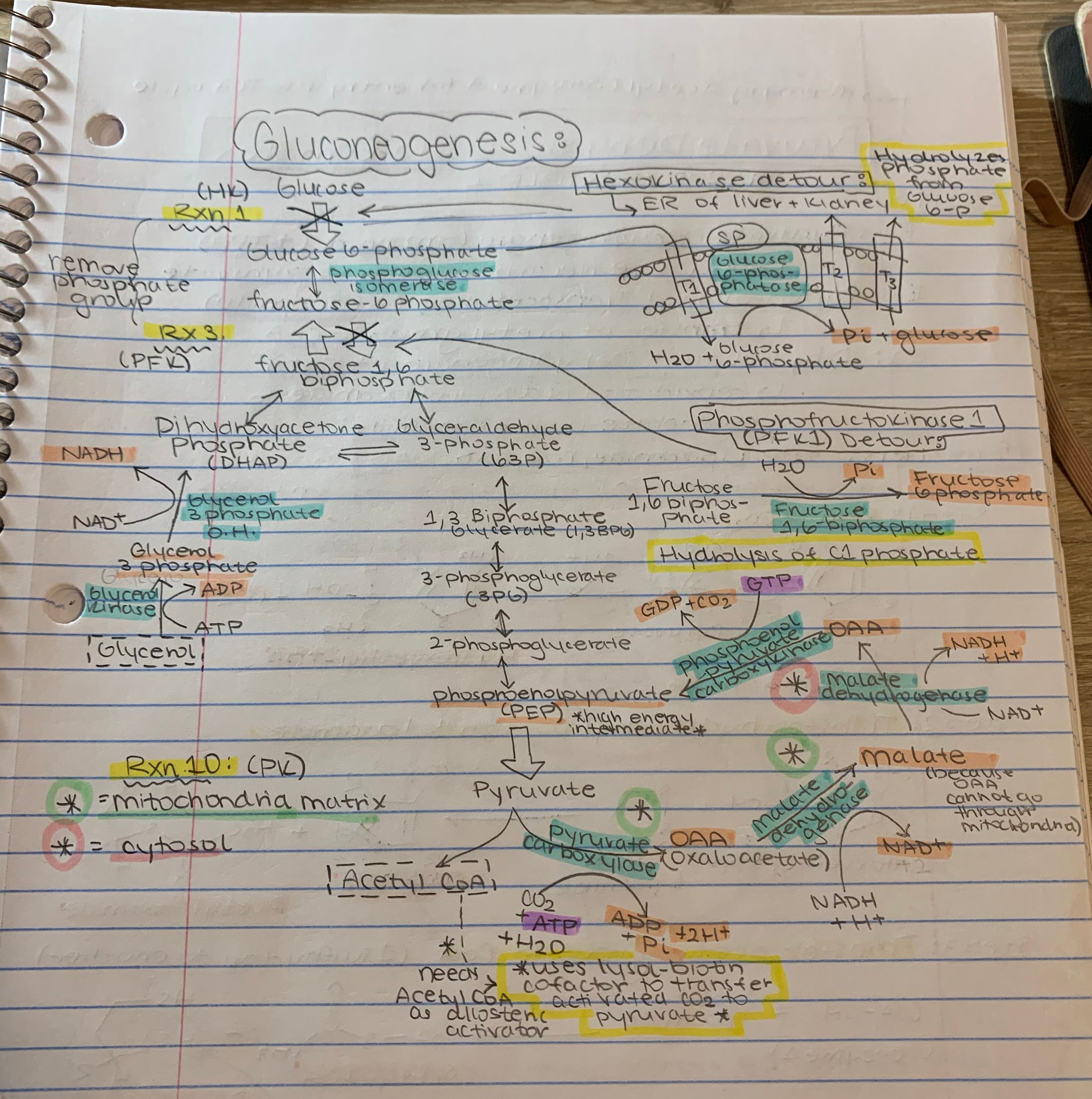
“I'm a visually oriented person. I see and learn medicine best through this perspective,” Herman noted. “Through the BLD program and medical school, I’ve learned the intricate structures and functions of the body by drawing out the various physiological and biochemical pathways, the microstructure of organs, and the complex cellular processes that make life possible."
“I like to draw things out with arrows, and I like to color code my notes," said Vezina. "And if there are different morphologies that I need to know in the lab between certain red cells or certain white cells, I like to draw them out on my own.”
This connection runs deep. As Herman and Vezina continue to grow, they further intertwine their passions for art and laboratory sciences; strengthening their knowledge while promoting the important impact medical laboratory scientists have on patient and public health. For laboratory medicine impacts the physical, art impacts the soul.
Banner image: One of Meredith Herman’s recent paintings, “Life Magnified,” which was inspired by her experiences in BLD and the pathology fellowship, was highly commended by the Royal College of Pathologists during its annual international art competition, the Art of Pathology.

